What is ultrasonography? What does it do?
Ultrasonography works with sound waves and does not contain radiation. The examination can be performed with the patient lying in the supine position or lying on his side. During the examination, the gel is applied and no pain is felt. It can also be used safely in pregnant women. In young women, in the presence of dense breasts, in the evaluation of lesions detected on mammography, ultrasonography examination is performed in fibrocystic breast structure. The main examination method for breast in women and children under 40 years of age is ultrasonography.
However, it cannot be used as a substitute for mammography in women over the age of 40 and in cancer screening. It is added to mammography as an adjunct method.
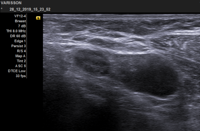
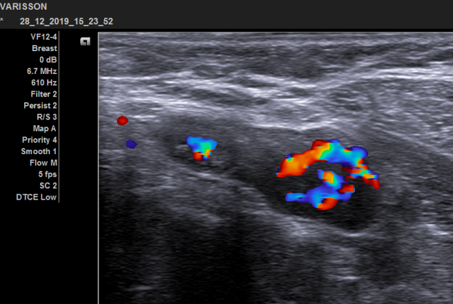
If any suspicious lesion is detected during the examination, its edge features, shape, and internal structure are examined and simultaneous Doppler (Color) Ultrasound is used to evaluate the vessel content. In addition, it is possible to perform a biopsy by seeing the lesion under ultrasound guidance in suspicious lesions.
Who is ultrasonography performed for?
- Children with breast enlargement or lesions
- For men with breast enlargement or a palpable mass
- For pregnant and lactating
- women under the age of 40
- Women with dense breast tissue and fibrocystic breast pattern
- Those who have signs of infection such as redness, swelling, pain in the breast
- Those with suspicious findings on physical examination or mammography
- In the examination of the underarm lymph nodes
- It is used as a guide method in biopsy in patients with lesions detected in the breast.
How many minutes does the test take?
Ultrasonography examination takes 20-60 minutes, depending on the condition of the patient.
What to do before the inspection?
No special preparation is required for ultrasonography examination.
What are the points to be considered after the examination?
There is nothing to pay special attention to after the examination. The information and findings obtained after the examination are documented as a report and presented to the patient/relevant doctor.
**** Don’t forget to bring all your old breast tests with you when you come to breast ultrasound, mammography or biopsy procedures!!!
