What is ultrasonography? What does it do?
Ultrasonography (US) is an imaging method that does not contain radiation, image is obtained through sound waves and is used in the diagnosis of many diseases. It is not a method that causes side effects, pain or suffering. It is the first preferred imaging method in pregnant and children. It can be used in imaging of internal organs in the abdomen, musculoskeletal system diseases, pregnancy follow-up during pregnancy, pediatric patients, breast diseases, etc. It is a safe method first used in many cases. In addition to the superficial thyroid, lymph node, testis, muscle and tendon structures and joints, it is possible to visualize parts of the body that cannot be accessed by endoscopy with ultrasonography. Ultrasound is used in the diagnosis, follow-up and control processes of diseases in these areas. When biopsy procedures are performed under ultrasound guidance, since pathological tissue is seen and the procedure is performed, it will be easier to reach the correct diagnosis and biopsy repetitions can be prevented. There is a chance to obtain real-time and dynamic images.
What are the Usage Areas of Ultrasonography?
Ultrasonography is the imaging method of first choice in children. It is used in the follow-up and control of both the mother and the baby in pregnancy follow-up. It is the first imaging method to be used in pregnant and lactating women. In the investigation of diseases of the gallbladder, liver, pancreas, in the evaluation of gallbladder and kidney stones, in the evaluation of tumoral or infectious diseases of breast, armpit and other soft tissue and superficial tissues, in the evaluation of muscle tendons and joints, in the size and diseases of the thyroid, testicular, ovary, uterus and It is used as a guide method in the evaluation of diseases of organs such as prostate and in interventions such as cysts and biopsy. In addition, it can guide methods such as laser, microwave, cryoablation in treatment.
How many minutes does the test take?
Ultrasonography examination takes an average of 20-60 minutes, depending on the type of organ to be examined or the area to be examined.
What to do before the inspection?
No special preparation is required for ultrasonography examination. In the ultrasonography to be performed for the lower abdomen, slightly congested urine will facilitate imaging. In ultrasonography for the upper abdomen, fasting for 4-6 hours will provide better visualization of organs such as pancreas and gall bladder.
What are the points to be considered after the examination?
There is nothing to pay special attention to after the examination. The information and findings obtained after the examination are documented as a report and presented to the patient/relevant doctor.
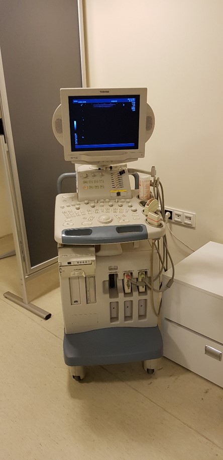
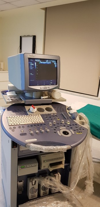
* There are 3 Doppler Ultrasonography/Doppler Ultrasonography devices (SIEMENS, Toshiba and General Electric) in our department.
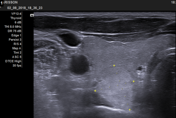
Ultrasonography image of the nodule located in the posterior part of the right thyroid lobe 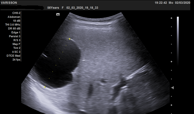
Ultrasonography image of cystic mass lesion in the spleen
